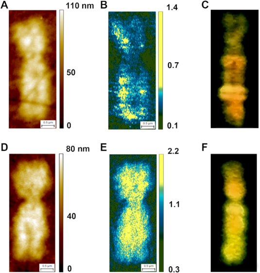
Figure 5.

AFM-IR on Xs female chromosomes: (A and D) AFM topography of active X (A) and inactive—methylated X (D) female chromosome. (B and E) The ratio of the integrated absorption band at 2952 cm−1 to the absorption band at 1240 cm−1 of active X (B) and inactive X (E) female chromosome. (C and F) anty 5-methylcytosine direct immunostaining counterstained with propidium iodide of active X (C) and inactive X (F) female chromosome, similarities between B, C and E, F can be observed; chromosome thickness Xa 65 – 80 nm, Xi 60 – 80 nm, pixel sizes Xa 7.5 × 14 nm, Xi 6.2 × 14 nm.
