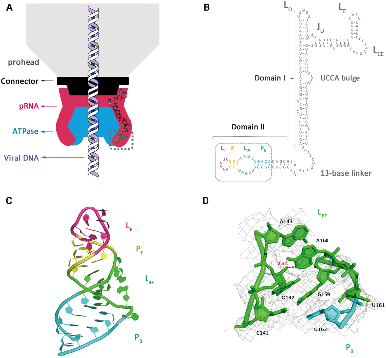
Figure 1.
Crystal structure of phi29 pRNA domain II. (A) Schematic of bacteriophage phi29 DNA packaging motor. The viral capsid, connector, pRNA and ATPase are shown in gray, black, magenta and blue, respectively. Model of the pRNA domain I was reconstructed with previously published structures (PDB accession codes: 3R4F, 4KZ2). Viral DNA is shown in the middle. Gray dashed box labeled the crystallized pRNA domain II in this study. (B) Sequence and secondary structure prediction of the pRNA. The PB, LBF, PF and LF are colored in cyan, green, yellow and magenta, respectively. The crystallization construct in this study is labeled by the gray dashed box. (C) Crystal structure of the pRNA domain II (PDB code: 6JXM). (D) A close-up view of the LBF loop superposed on 2Fo-Fc electron density, contoured at 1.0 sigma. N1 of the A160 makes a hydrogen bond to the 2′-OH of G142. Distance is given in angstrom in red.

