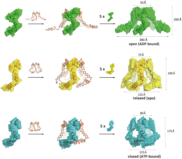
Figure 4.
Reconstructed pentameric 3D models of ADP-, apo- and ATP-bound RNP. The full-length pRNA structure was docked into previously published cryo-EM and atomic force microscopy structure to reconstruct a pentameric framework (23). Then the ADP–bound RNP SAXS bead model (green), RNP SAXS bead model (yellow) and ATP–bound RNP SAXS bead model (cyan) were fitted onto the pentameric framework. The top inner pore, bottom inner pore and height of the ‘open’ model are ∼65, 380 and 200 Å. The top inner pore, bottom inner pore and height of RNP pentameric model are ∼70, 155 and 190 Å. The top inner pore, bottom inner pore and height of the ATP–bound RNP pentameric model are ∼80, 115 and 175 Å. The lower arm angle difference between the ATP and ADP-bound complex is ∼93°.

