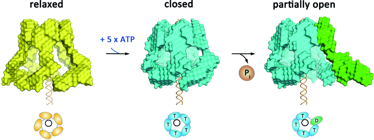
Figure 5.
Reconstructed 3D models of the phi29 molecular movement during ATP binding and hydrolysis. During ATP binding, the individual RNP switches from the ‘relaxed’ conformation (yellow bead models) to the ‘closed’ conformation (blue bead model labeled with ‘T’s). Subsequent ATP hydrolysis opens the complex (green subunit labeled with ‘D’) by swinging the pRNA domain II outwards.

