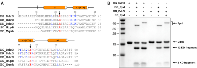
Figure 1.
Protein characterization and sequence alignment. (A) Structural based sequence alignment of DNA binding domains of XRE family proteins. DdrO proteins from D. geothermalis, D. radiodurans and D. deserti are denoted by DG-DdrO, DR-DdrO and DD-DdrO, respectively. HipB from S. oneidensis and MqsA from E. coli are denoted by SO-HipB and EC-MqsA, respectively. Residues conserved in XRE family proteins and DdrO proteins are highlighted in red and blue, respectively. The black arrowheads indicate the signature RE pair of XRE family proteins. (B) SDS-PAGE gel showing the DdrO cleavage by PprI. For the reaction, 8 μM of DG-DdrO or DR-DdrO was incubated with 1 μM of DG-PprI or DR-PprI in the presence of Mn2+ at 37°C for 30 min. DdrO, PprI and two product fragments are indicated by black arrowheads.

