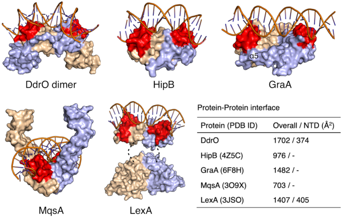
Figure 3.
Structural comparison of the dimerization between DG-DdrO and XRE family proteins. DG-DdrO, HipB (PDB ID: 4Z5C), MqsA (PDB ID: 3O9X), GraA (PDB ID: 6FIX) and LexA (PDB ID: 3JSO) are shown as surface and two protomers are colored in wheat and light blue, respectively. The HTH motifs are highlighted in red. For DdrO protein, DNA from MqsA-DNA complex was docked onto the DG-DdrO protein by superposition between HTH motifs of DG-DdrO and MqsA. The position of α5 in GraA is labeled. The disordered link regions in LexA are indicated by dashed lines. The dimeric interface between two protomers were calculated by PDBePISA (37) and listed in the table.

