
Figure 4.
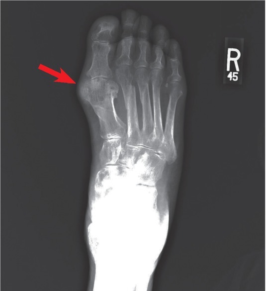
Severe gout presenting on the first metatarsophalangeal joint. AP X-ray of the right foot shows a medial pararticular calcified soft tissue mass at the level of the first metatarsophalangeal joint (red arrow), resulting in adjacent intraosseous erosions with sclerotic borders.