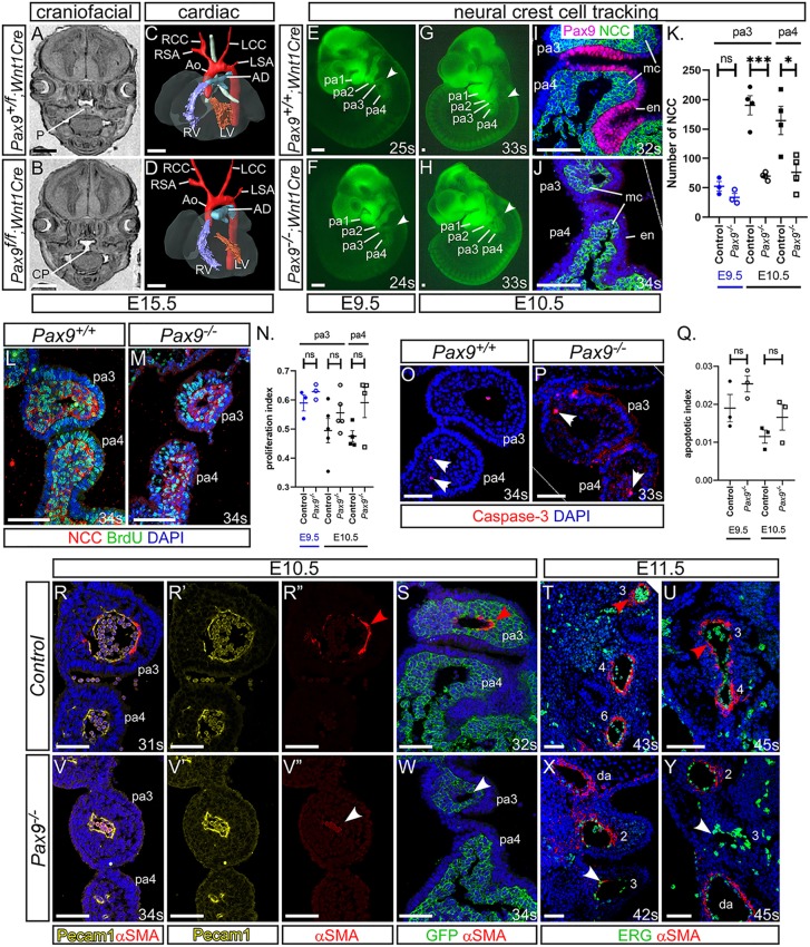
Fig. 4.
Failure in smooth muscle cell recruitment causes the 3rd PAAs to collapse. (A-D) E15.5 embryos with a Pax9 conditional deletion in neural crest cells (NCCs) were examined by MRI. (A) Control embryos had a normal palate (P). (B) Pax9f/f;Wnt1Cre mutant embryos (n=6) had cleft palate (CP). (C,D) No cardiovascular defects were observed in control or mutant embryos. Ao, aorta; AD, arterial duct; LCC, left common carotid artery; LSA, left subclavian artery; LV, left ventricle; RCC, right common carotid artery; RSA, right subclavian artery; RV, right ventricle. (E-J) NCCs in Pax9+/+ (E,G,I) or Pax9–/– (F,H,J) embryos were labelled using Wnt1Cre and eYFP alleles. (E-H) Fluorescence imaging showed no difference in NCC migration into the pharyngeal arches (arrowheads) at E9.5 (n≥3, 23-25 s) and E10.5 (n≥5, 32-36 s). (I,J) Fluorescent embryos were sectioned coronally and immunostained using anti-GFP and anti-Pax9 antibodies. (K) NCCs were counted from immunostained sections. *P<0.05, ***P<0.001 (two-tailed unpaired t-test). (L,M) Embryo sections were immunostained with anti-BrdU and anti-GFP antibodies to detect proliferation in NCCs in Pax9+/+ (L) and Pax9–/– (M) embryos. (N) No significant difference in the rate of proliferation was found between control and Pax9–/– NCCs in the caudal arches at E9.5 (n=3, 23-28 s) or E10.5 (n≥4, 31-39 s). (O,P) Embryo sections immunostained using an anti-caspase 3 antibody to detect apoptosis in the caudal pharyngeal arches of Pax9+/+ (N,O) and Pax9–/– (P) embryos. (Q) No significant difference in the rate of apoptosis was found between control and Pax9–/– embryos at E9.5 (n=3, 24-28 s) or E10.5 (n=3, 30-35 s). Two-tailed unpaired t-test. (R-Y) Embryo sections were immunostained using anti-αSMA antibody for smooth muscle (R,R″-U,V,V″-Y,S,W), anti-Pecam1 (R,R′,V,V′) or -ERG (T,U,X,Y) antibodies for endothelium, or anti-GFP to label NCCs in Wnt1Cre;eYFP embryos (S,W). In all control embryos, SMC surrounded the 3rd PAAs [E10.5, n=7, 31-36 s (R,S); and E11.5, n=3, 43-45 s (T,U); red arrowhead]. In Pax9–/– embryos, the 3rd PAAs had limited recruitment of SMCs [E10.5, n=6; 32-35 s (V,W); and E11.5, n=3, 42-45 s (X,Y); white arrowheads]. da, dorsal aorta; en, endoderm; mc, mesenchyme; ns, not significant; pa, pharyngeal arch. Somite counts (s) are indicated. Scale bars: 500 μm in A-D; 100 μm in E-J,L,M,O,P; 50 µm in R-Y. The somite numbers given in the legend reflect the range analysed for the whole study. The figure contains representative images only.

