Fig. 6.
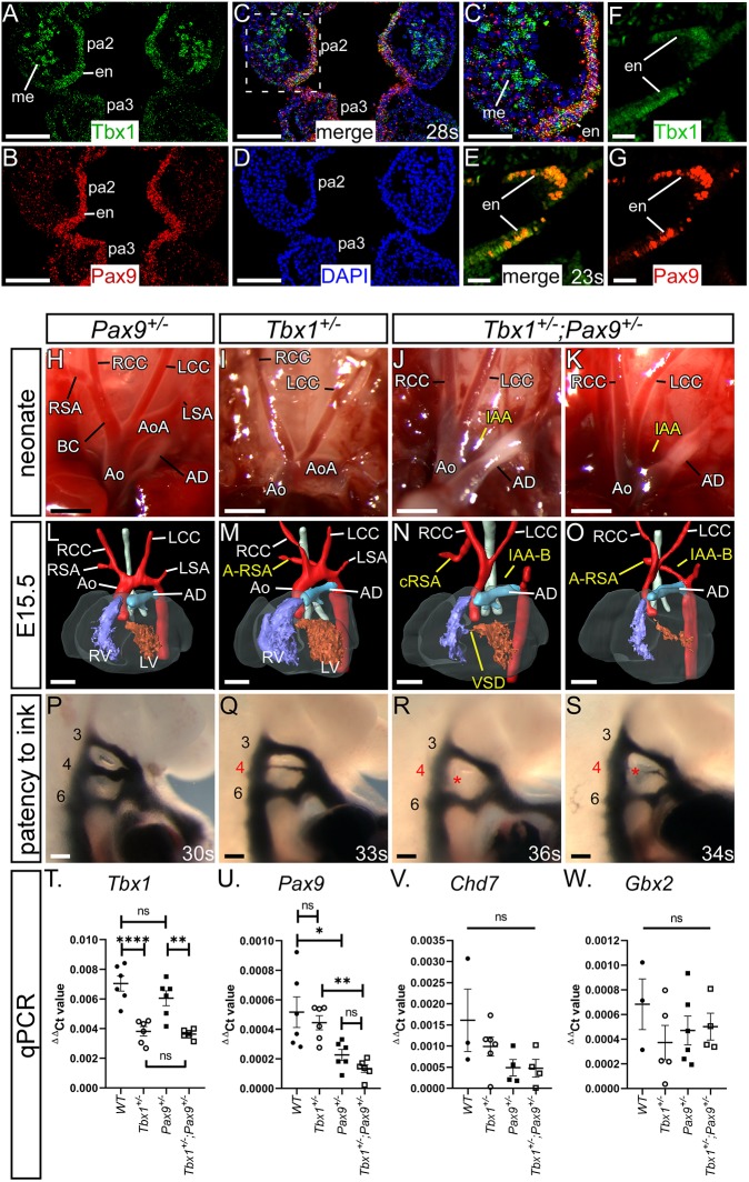
Tbx1;Pax9 double heterozygous embryos have 4th PAA defects. Tbx1 and Pax9 expression in the pharyngeal arches was examined at E9.5. (A-D) RNAScope in situ hybridisation expression of Tbx1 in the 2nd and 3rd pharyngeal arch (pa), endoderm (en) and mesoderm (me; A), and Pax9 expression in the endoderm only (B). The signals overlap in the endoderm (C, and higher power in C′). (D) DAPI staining. (E-G) Immunostaining for Tbx1 and Pax9 (E) demonstrates protein colocalisation within cells of the pharyngeal endoderm. Single staining for Tbx1 (F) and Pax9 (G) is shown. (H-K) Neonates from a Tbx1+/– and Pax9+/– intercross were recovered and the aortic arches examined. (H) Arch arteries were normal in all Pax9+/– neonates (n=10). (I) Tbx1+/– neonates (n=6) often presented with A-RSA, inferred by the absence of the brachiocephalic and right subclavian arteries. (J,K) All Tbx1+/–; Pax9+/– neonates (n=9) died within 24 h of birth with IAA and/or A-RSA. (L-O) 3D reconstructions of E15.5 embryo hearts from MRI datasets. (L) Pax9+/– hearts were normal (n=15). (M) Tbx1+/– hearts (n=19) frequently displayed 4th PAA defects such as A-RSA. (N,O) All Tbx1+/–; Pax9+/– embryos (n=20) presented with a 4th PAA-derived defect such as IAA-B, A-RSA and/or cervical right subclavian artery (cRSA). (P-S) Intracardiac ink injections into E10.5 embryos (27-38 s). (P) PAAs in control embryos were normal (n=10). (Q) In Tbx1+/– embryos (n=8) the 4th PAAs were often hypoplastic. (R,S) In Tbx1+/–;Pax9+/– embryos (n=9), the 4th PAAs were frequently bilaterally absent. Ao, aorta; AoA, aortic arch; AD, arterial duct; BC, brachiocephalic; LCC, left common carotid artery; LSA, left subclavian artery; LV, left ventricle; RCC, right common carotid artery; RSA, right subclavian artery; RV, right ventricle. Somite counts (s) are indicated. Scale bars: 2 mm in H-K; 500 μm in L-O; 100 μm in A-D,P-S; 50 µm in E-G. (T-W) qPCR analysis in wild-type, Tbx1+/–, Pax9+/– and Tbx1+/–;Pax9+/– embryos (n≥3 per genotype, somite range 23-27). (T) Tbx1 levels were significantly reduced in Tbx1+/– and Tbx1+/–;Pax9+/– embryos. (U) Pax9 levels were significantly reduced in Pax9+/– and Tbx1+/–;Pax9+/– embryos. There was no significant difference in Tbx1 (T), Pax9 (U), Chd7 (V) or Gbx2 (W) levels between single heterozygotes and Tbx1+/–;Pax9+/– embryos. One-way ANOVA with Tukey's multiple comparison test. ns, not significant. *P<0.05, **P<0.01, ****P<0.0001.

