Fig. 7.
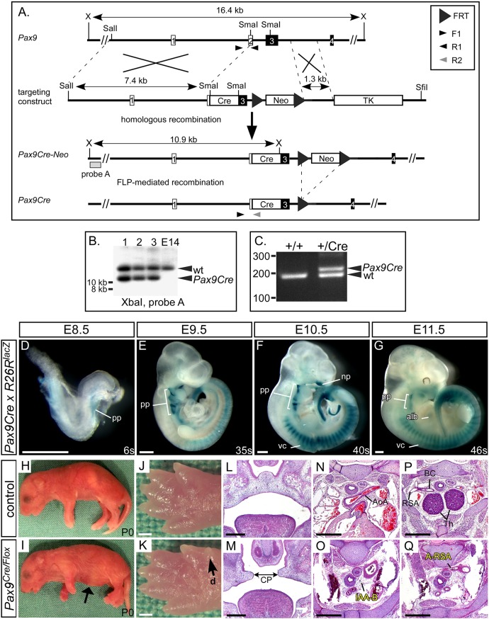
Generation of the Pax9Cre mouse. (A) A Pax9Cre targeting vector was created with a Cre cassette inserted into the SmaI restriction sites found in exons 2 and 3 of the Pax9 gene. For transformation, the vector was linearised with SfiI. (B) Correctly targeted ES cells were identified by Southern blotting on XbaI-digested genomic DNA. (C) The derived Pax9Cre-Neo mice were crossed with FLPe-expressing mice to remove the neomycin cassette and create Pax9Cre mice, which were genotyped using the indicated primers. (D-G) Pax9Cre mice were mated with R26RlacZ reporter mice and embryos were stained with X-Gal at E8.5 (n=3), E9.5 (n=7), E10.5 (n=6) and E11.5 (n=5). Positive staining in regions known to express Pax9 was observed (alb, anterior limb bud; np, nasal process; pp, pharyngeal pouch; vc, vertebral column). (H-Q) Pax9Cre and Pax9flox/flox mice were crossed to create Pax9+/flox (control) and Pax9Cre/flox (mutant) mice. All Pax9Cre/flox neonates died on the day of birth and presented with the typical Pax9-null phenotype (n=5), including a bloated abdomen (I, arrow) and pre-axial digit duplication (d) in the hind limb (K, arrow). Sections of E14.5-E15.5 embryos (n=6) showed cleft palate (CP; M), absent thymus and cardiovascular defects (O,Q). AoA, aortic arch; BC, brachiocephalic; Th, thymus. Somite counts (s) are indicated. Scale bars: 500 µm.

