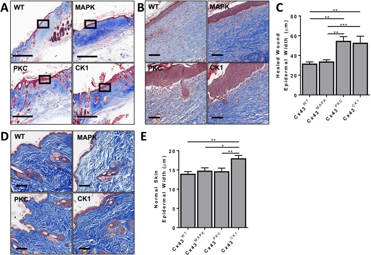
Fig. 2.
Cx43PKC and Cx43CK1 have thicker skin epidermis after reepithelization of wounds. (A,B) Representative immunohistochemical images of Masson's Trichrome staining in the healed wounds of Cx43WT, Cx43MAPK, Cx43PKC and Cx43CK1 mice. Black boxes represent the origin of the magnified image shown in B. (C) Quantification of the epidermal thickness in healed wounds. n=4 (43) Cx43WT, 5 (96) Cx43MAPK, 3 (27) Cx43PKC, and 5 (54) Cx43CK1 mice (sites) per group. (D) Representative immunohistochemical images of Masson's Trichrome staining in the skin of Cx43WT, Cx43MAPK, Cx43PKC and Cx43CK1 mice. (E) Quantification of the epidermal thickness in normal skin. n=4 (55) Cx43WT, 5 (58) Cx43MAPK, 5 (62) Cx43PKC, and 6 (82) Cx43CK1 mice (sites) per group. Results are mean±s.e.m., *P<0.01, **P<0.005, ***P<0.0005 (one-way ANOVA). Scale bars: 500 µm (A,D), 100 µm (B).

