Fig. 3.
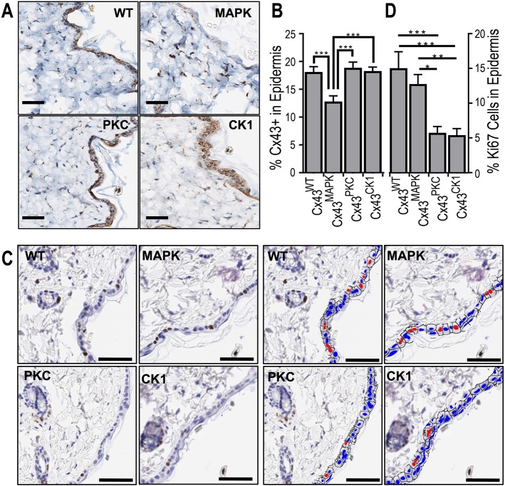
Cx43MAPK mice have reduced Cx43 expression and Cx43PKC mice have decreased proliferation in the epidermis. (A) Representative immunohistochemical images of Cx43 expression in the skin of Cx43WT, Cx43MAPK, Cx43PKC and Cx43CK1 mice. (B) Quantification of Cx43 expression by immunohistochemistry in the epidermal layer of mouse skin. n=8 (66) Cx43WT, 9 (44) Cx43MAPK, 10 (49) Cx43PKC, and 10 (51) Cx43CK1 mice (sites) per group. (C) Representative immunohistochemical images of Ki67 expression in the skin of Cx43WT, Cx43MAPK, Cx43PKC and Cx43CK1 mice. Right and left quad-panels are identical except the right is pseudocolored (red, Ki67+; blue, Ki67−) for visual clarity. (D) Quantification of Ki67 expression by immunohistochemistry in the epidermal layer of mouse skin. n=8 (50) Cx43WT, 9 (44) Cx43MAPK, 10 (50) Cx43PKC, and 10 (50) Cx43CK1 mice (sites) per group. Results are mean±s.e.m. *P<0.05, **P<0.005, ***P<0.0005 (one-way ANOVA). Scale bars: 100 µm.

