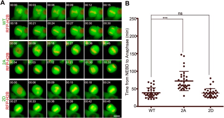
Fig. 5.
Phosphorylation of importin-α1 at T9 and S62 is required for mitotic progression. (A) HeLa cells were co-transfected with GFP–importin-α1 WT or mutants (2A and 2D) and RFP–H2B (as a chromatin marker) and then were subjected to automated time-lapse live-cell fluorescence imaging. The GFP signals indicate cells transfected with GFP importin-α1 or mutants. The red signals indicate H2B. The onset of NEBD is marked as 0 min. Scale bar: 10 µm. (B) Statistics showed that the average time from NEBD to anaphase was prolonged by non-phosphorylation-mimicking mutant GFP-importin-α1 2A. Results are mean±s.d., n=50 cells per group. ***P<0.001; ns, not significant (Student's t-test).

