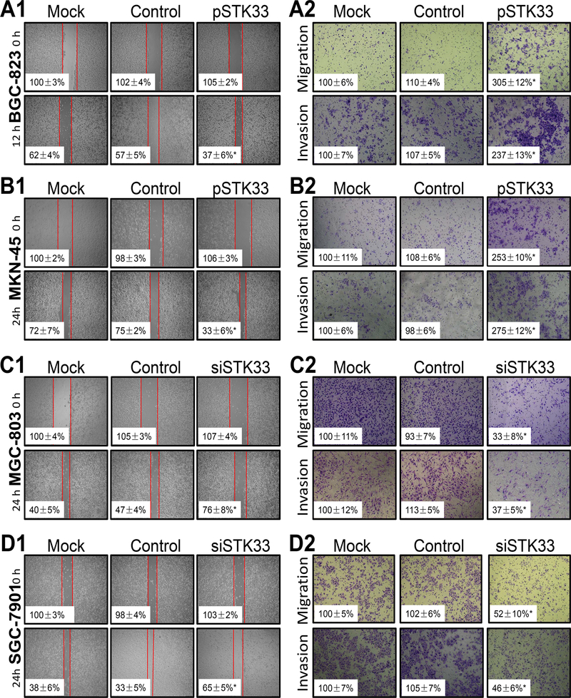
Figure 2. Influence of STK33 expression on gastric cancer cell migration and invasion in vitro.
BCG-823 (A1 and A2), MKN-45 (B1 and B2), MGC-803 (C1 and C2), and SGC-7901 (D1 and D2) cells were transfected with pSTK33, siSTK33, control vectors, or siRNAs (mock) for 48 hours. For a cell scratch-wound assay, cells in each group were placed in six-well plates, wounded via scratching, and maintained at 37°C for at least 12 hours. Cell cultures were photographed, and cell migration was assessed by measuring the cell-free areas in multiple fields (the insert numbers indicate the percentage mean gap areas ± standard deviation in triplicate). The migration and invasion of gastric cancer cells were determined as described in Materials and Methods. The data represent the means ± standard error of the mean in triplicate from one representative experiment of three with similar results. *P < 0.05 in comparisons of pSTK33-treated, siSTK33-treated, mock, and control groups (Student t-test).

