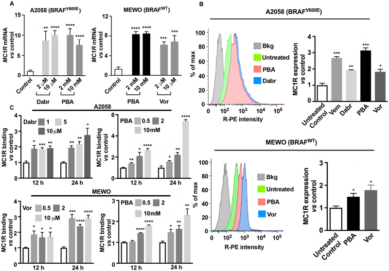
Figure 3.
MC1R expression is upregulated in human melanoma cells upon treatment with BRAFi and HDACi. (A) qRT-PCR analysis of MC1R mRNA expression in BRAFV600E A2058 cells after 24 h of incubation with BRAFi dabrafenib (Dabr) and HDACi 4-phenylbutyrate (PBA) and in BRAFWT MEWO cells after 24 h treatment with HDACi PBA and vorinostat (Vor) (n = 3). Data are presented as normalized mean MC1R mRNA ± SEM; *p < 0.05, **p < 0.01, ***p < 0.001, and ****p < 0.0001 vs controls; (B) flow cytometry histograms and protein expression of MC1R in A2058 cells after 24 h treatment with BRAFi: Vem (5 μM) and Dabr (2 μM); HDACi: PBA (2 mM) and Vor (2 μM); and in MEWO cells after incubation with PBA (2 mM) and Vor (2 μM). Experiments were conducted in triplicate. Data are expressed as relative expression of MC1R vs isotype control (mean ± SEM; *p < 0.05, **p < 0.01, ***p < 0.001 vs controls); (C) MC1R binding with [125I]NDP-α-MSH in A2058 and MEWO cells following incubation with Dabr (1−10 μM), Vor (0.5−10 μM), and PBA (0.5−10 mM) for 12−24 h (n = 4). Data are expressed as MC1R-ligand binding vs dimethyl sulfoxide (DMSO)-treated cells (mean ± SEM; *p < 0.05, **p < 0.01, ***p < 0.001, ****p < 0.0001 vs controls); all experiments were performed in duplicate (n = 2).

