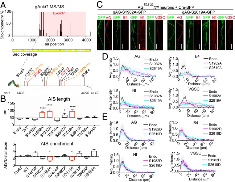
Fig. 4.
Phosphorylation of gAnkG at S1982 and S2619 is required for recruitment of β4-spectrin to the AIS. (A) MS-MS detected multiple high stoichiometry phosphorylation sites (>10%) in exon 37-encoded sequence from AnkG isolated from mouse brain by immunoprecipitation (SI Appendix, SI Materials and Methods). Locations of screened phosphorylation sites (black and red) and human neurodevelopmental disorder mutations (yellow) in gAnkG. Asterisk-labeled site S2406 corresponds to S2417 in mouse sequence. (B) Hippocampal neurons from AGE22−23fl/fl mice were cotransfected on day 3 with plasmids encoding Cre-BFP and gAnkG-GFP bearing serine/threonine to alanine mutants at indicated amino acid sites. Neurons were fixed and stained with AnkG antibody at 7 DIV. The length and the enrichment of AnkG at AIS is quantified. Mean ± SEM; *P < 0.05; ****P = 0.0001; 1-way ANOVA followed by Dunnett’s multiple comparisons test; n = 10 from 3 independent experiments. (C) AGE22−23fl/fl neurons transfected with plasmids encoding Cre-BFP and nonphosphorylatable gAnkG mutants (gAG-S1982A-GFP or gAG-S2619A-GFP) were stained with indicated antibody. (Scale bar, 20 μm.) (D) Average intensity of indicated antibody at the AIS is plotted (S1982A in magenta; S2619A in blue) and aligned with nontransfected neurons (endo, black line). n = 10 of each plot. Results were repeated in 3 independent experiments. (E) Average intensity of indicated antibody at the AIS is plotted for AGE22−23fl/fl neurons cotransfected with plasmids encoding Cre-BFP and phosphomimetic gAnkG mutants (S1982D in magenta or S2619D in blue) and aligned with nontransfected neurons (endo, black line). n = 10 of each plot. Results are repeated in 3 independent experiments.

