Fig. 4.
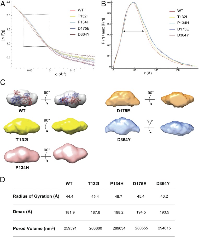
SAXS analysis of WT HisRS and its CMT disease mutants. (A) Experimental solution scattering data for the HisRS proteins. The dotted square shows the medium-resolution region of the scattering spectra. (B) P(r) of the SAXS data determined during data reduction with GNOM. (C) Two different views of the ab initio envelopes generated with DAMMIF for the HisRS proteins. The structure of the dimeric HisRS was manually docked into the envelope of HisRSWT. (D) Summary table of main SAXS parameters for the proteins tested.

