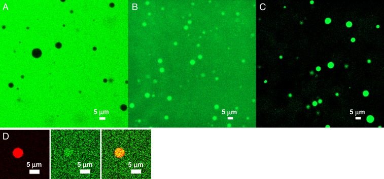
Fig. 4.
Confocal images of 3 FITC-labeled regulators, revealing different extents of recruitment into SH35–PRM5 droplets formed by equimolar mixing of the 2 proteins at 40 µM: (A) 5 µM FITC–Ficoll 70 plus 200 g/L Ficoll 70; (B) 2.5 µM FITC–lysozyme; (C) 2.5 µM FITC–heparin; (D) 1 µM Alexa 594–SH35 plus 1 µM fluorescein-sodium salt, showing enrichment of SH35 in the droplet phase (Left, red channel; similar to Fig. 2A) and nearly equal partitions of free fluorescein label in the 2 phases (Middle, green channel). The merge of the 2 channels is shown on the Right.

