Fig. 4.
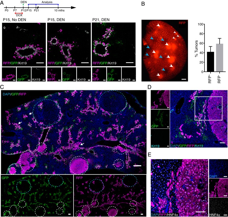
Lgr5+ hepatocytes are the primary cellular origin in DEN-induced HCC. (A) Experimental strategy for doxycycline (DOX) and DEN induction for Lgr5-rtTA/TetO-Cre/R26-tdTomato mice. Representative confocal images showing distribution of Lgr5-GFP+tdTomato+ around central veins at P15 without DEN (25 mg/kg) injection, and at P15 and P21, which were 3 d and 9 d after DEN injection, respectively. Dams were treated with doxycycline at P7–P12 of their litters to induce the labeling of tdTomato in Lgr5-expressing hepatocytes of young male pups. (B) Representative whole-mount fluorescence view of a tumorous liver lobe of Lgr5-rtTA/TetO-Cre/R26-tdTomato mouse harvested at 8 mo after DEN induction. tdTomato+ and tdTomato− tumors are depicted by white and blue arrowheads, respectively. Approximately 40% of the tumors detected in the liver of triple transgenic mice showed native tdTomato+ (RFP+) fluorescence. n = 4 mice. (C) Representative confocal tile scan images of a tumorous liver lobe harvested from DEN-treated Lgr5-rtTA/TetO-Cre/R26-tdTomato mouse showing tdTomato+ (white circles) and tdTomato− (blue circles) tumors. White arrows indicate possible tdTomato+ tumor initiation sites. All DEN-induced tumors were negative for GFP. n = 4 mice. (D and E) Representative confocal images showing that tdTomato+ tumors are GFP and Krt19-negative (D), and HNF4α-positive (E). (Scale bars: B and C, 200 μm; D and E, 100 μm.)

