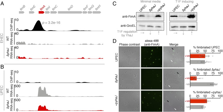
Fig. 4.
YhaJ enhances expression of type 1 fimbriae in UPEC. (A) Expanded view of the YhaJ binding site upstream of the silent T1F locus in EHEC identified by ChIP-seq. The associated RNA-seq tracks highlight the lack of transcription from this silent locus in both WT EHEC (gray) and ΔyhaJ (red). (B) ChIP-seq data from the active T1F locus in UPEC illustrating the lack of YhaJ enrichment but a downshift in fimA transcription for ΔyhaJ (red) versus WT UPEC (gray). (C) Immunoblot analysis of FimA expression levels from WT UPEC, ΔyhaJ, and the complemented mutant +pyhaJ grown in minimal media (ChIP/RNA-seq conditions) and T1F-inducing conditions (static LB at 37 °C). Levels of GroEL were used to assess equal loading, and the influence of yhaJ mutation/complementation on T1F expression is highlighted (Bottom). Multiple biological replicates of immunoblots were performed. (D) Phase-contrast microscopy of WT UPEC, ΔyhaJ, and +pyhaJ grown under T1F-inducing conditions overlaid with immunofluorescence images of the cells probed using anti-FimA and Alexa-488 antibodies. The level of fimbriation around single cells was assessed (black arrows, T1F-positive; white arrows, T1F-negative) from more than 5 random fields of view per replicate and expressed as percentage fimbriated cells within the population (Right). Error bars represent SD, and the experiments were performed on 3 independent occasions.

