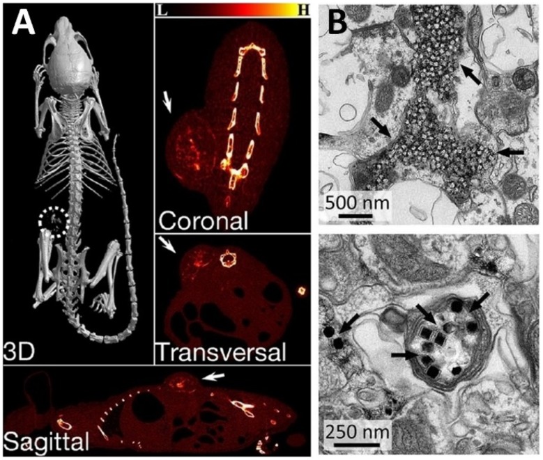
Figure 10.
(A) X-ray CT images of the tumor-bearing mice after intratumoral injection of NP/PDA suspension. Reproduced with permission from Dai Y, Yang D, Yu D, et al. Mussel-inspired polydopamine-coated lanthanide nanoparticles for NIR-II/CT dual imaging and photothermal therapy. ACS Appl Mater Interfaces. 2017;9(32):26674– 26683.186 Copyright American chemical society 2017. (B) Electron micrographs of UCNPs distributed in the neuronal tissue. Black arrows indicate clusters of UCNPs. Top image shows the distribution of most UCNPs in extracellular space, and the bottom image shows the uptake of UCNPs within an axon. From Chen S, Weitemier AZ, Zeng X, et al. Near-infrared deep brain stimulation via upconversion nanoparticle–mediated optogenetics. Science. 2018;359(6376):679.187 Reprinted with permission from AAAS https://science.sciencemag.org/content/359/6376/679.

