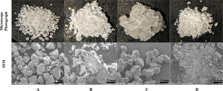
Figure 1.
Upper row: Microscope photographs, magnification 10x. Lower row: Scanning electron microscopy (SEM) of: (A) (+)-Xylitol, (B) Crystallized (+)-xylitol, (C) Physical mixture (PM), (D) OC-X 1:7 SD formulation. The SEM scale bar represents 1 mm. The magnification of A-D is: 40x, 45x, 30x, and 50x *1000, respectively.

