Figure 8.
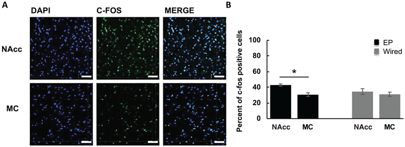
C-fos induced by EP and wired stimulation. (A) Representative confocal images (40×) of DAPI, c-fos, and merged DAPI and c-fos-stained images within the NAcc and MC in the left hemisphere (EP side) only; scale bar = 50 μm. (B) EP stimulation produced significantly more c-fos positive cells (shown here as a percentage of DAPI-stained cells) in the nucleus accumbens (NAcc), a brain region innervated by the MFB, compared to the motor cortex (MC), which does not have direct connections within the MFB. There was a slight increase in NAcc c-fos expression compared to MC with wired stimulation, but it was not statistically significant (n = 2, both animals received EP and wired stimulation). *p < 0.05. Error bars represent SEM.

