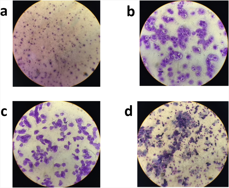
Fig. 2.
Representative images from each stage of estrous. A sterile micropipette was used to flush the vaginal cavity of female rats with distilled water (McLean et al., 2012). The collected sample was placed on a glass slide and allowed to dry at room temperature. The slides were then stained with crystal violet and examined under a microscope to determine the stage of estrous. The following guidelines were used to determine the stage of estrous: (a) diestrus (many leukocytes, occasional nucleated epithelial cells, no cornified cells); (b) proestrus (many nucleated epithelial cells, occasional cornified cells, and few leukocytes); (c) estrus (many cornified cells, few nucleated epithelial cells, no leukocytes); (d) metestrus (some cornified cells, nucleated epithelial cells, and leukocytes).

