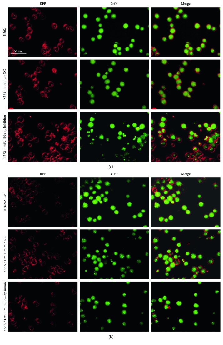
Figure 6.
After transfected with miR-199a-5p mimic and inhibitor in K562/ADM and K562 cells, respectively, the cells were subsequently infected with mRFP-GFP-LC3 adenovirus and were observed under a confocal fluorescence microscope. Representative images are presented to indicate the cellular localization patterns of mRFP-GFP-LC3 fusion protein.

