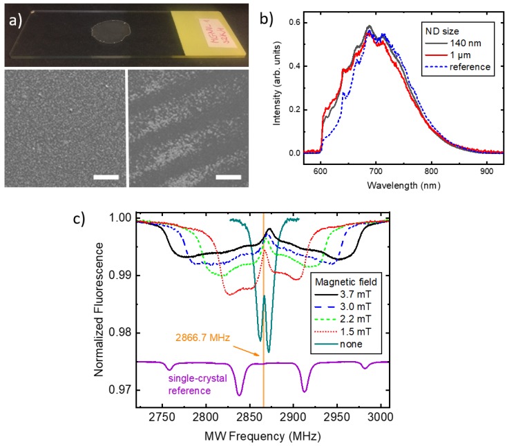
Figure 2.
Signals observed with deposited ND ensembles. (a) 1-m-size NDs deposited on a microscope slide (top panel) and SEM images of uniform (left panel) and self-organized (right panel) areas. The scale bar size is 100 m; (b) Optical fluorescence spectra of 140-nm and 1000-nm-size NDs, and a reference bulk type Ib diamond; (c) ODMR recorded with a dried 140-nm ND solution as a function of the magnetic-field strength. Vertical line indicates the symmetry frequency for the zero field. The reference signal was recorded with a [111]-oriented single-crystal in a 4 mT field (scaled-down by a factor of 10 and vertically offset for clarity).

