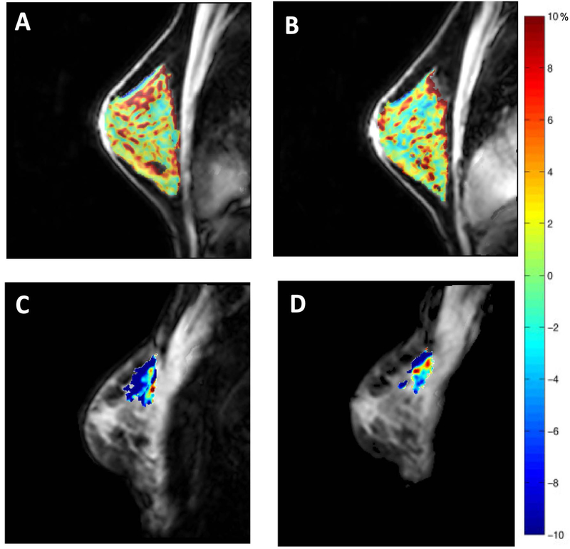
Figure 5.
Panels A and B display the repeatability of CEST measurements in the fibroglandular tissue of the healthy breast. Amide proton transfer residual maps are overlaid on corresponding anatomical images in the same subject before and after repositioning. Tumor amide proton transfer maps of a patient with breast cancer before (panel C) and after (panel D) one cycle of neoadjuvant chemotherapy. The patient achieved partial response to therapy.

