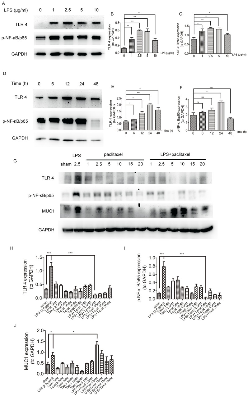
Figure 5.
Paclitaxel inhibited TLR 4-NF-κB/p65 activation and upregulated MUC1 in LPS-stimulated lung type II epithelial cell line. (A–C) A549 cells were stimulated with different doses of LPS (1, 2.5, 5, 10 μg/mL) for 24 hrs. (D–F) A549 cells were incubated with 2.5 μg/mg LPS for 6, 12, 24, and 48 hrs. (G–J) A549 cells were treated with different doses of paclitaxel (1, 2.5, 5, 10, 15, 20 nM) after stimulated by 2.5 μg/mL LPS for 24 hrs. TLR 4, p-NF-κB/p65 and MUC1 expression levels were detected by Western blot. Data represent means ± SD (n=6). *P<0.05, **P<0.01, ***P<0.001.

