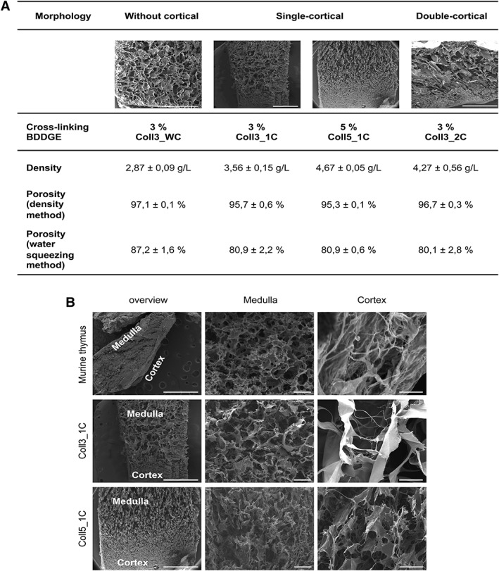
Figure 1.

Generation of collagen‐based scaffolds mimicking the thymic ultrastructure. (A): 3% and 5% 1,4‐butanediol diglycidyl ether (BDDGE) collagen type I scaffolds with different structures were tested to check their porosity, density, and perfusion ability. Scaffold dimension: 8 mm × 8 mm × 4 mm. (B): Electron microscopy images showing the ultrastructure of a murine native thymus and collagen type I scaffolds with two different percentages, 3% (Coll3_1C) and 5% (Coll5_1C), of the crosslinker BDDGE and one cortical layer. Our scaffolds recapitulate the medullary and cortical features of a normal thymus. Scale bar: 200 μm for medulla and cortex magnification; 10 mm for overview magnification.
