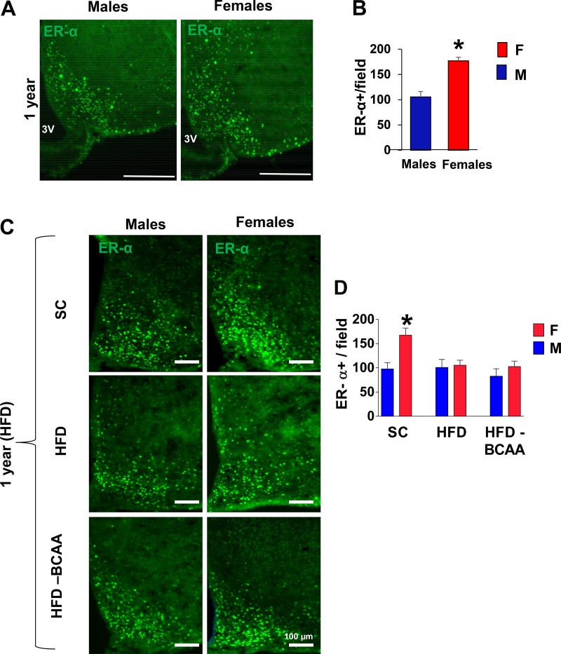
Fig. 5.
Estrogen receptor α (ERα) immunoreactivity in aged high-fat diet (HFD)-fed offspring. A: representative images of ER-α immunoreactivity in the mediobasal hypothalamus (MBH) subregion of 12-mo-old male and female mice. Scale bars: 100 µm. B: numbers of cells immunoreactive for ER-α in the arcuate nucleus of the hypothalamus (ARC) from 12-mo-old male and female mice (Student’s t-test). C: representative images of ER-α immunoreactivity in the ARC of 12-mo-old male and female offspring challenged with HFD for 2 wk. D: numbers of cells immunoreactive for ER-α in the ARC from male and female offspring of standard diet (SC), HFD, or HFD+BCAA dams (n = 6/group), error bars reflect means ± SE. *P < 0.01 vs. SC, as assessed by two-way ANOVA followed by Newman-Keuls test. BCAA, branched-chain amino acid.

