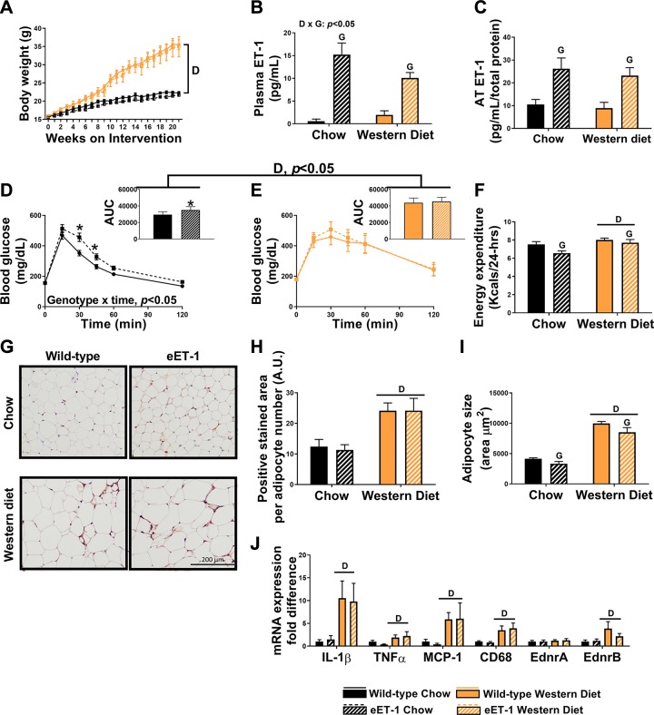
Fig. 1.
Overexpression of endothelin-1 (ET-1) impairs glucose tolerance but does not cause visceral adipose tissue (AT) inflammation in female mice. A: body weight growth curves over 21 wk of diet intervention. B: plasma ET-1 concentrations. C: ET-1 concentrations in perigonadal AT. D: glucose tolerance test (GTT) with area under the curve (AUC) conducted at 19 wk of intervention in chow-fed mice. E: GTT with an AUC at 19 wk of intervention in Western diet-fed mice. F: energy expenditure throughout 24 h. G: macrophage marker MAC-2 immunostained perigonadal adipose sections. Magnification ×20 with ×10 objective. H: average MAC-2-positive stained area. I: average adipocyte size per 100 cells. J: expression of inflammatory genes in perigonadal AT, represented as fold differences from wild-type chow. Values are means ± SE; n = 9–11/group. “D” indicates P < 0.05, main effect of diet. “G” indicates P < 0.05, main effect of genotype. “D × G” indicates P < 0.05, diet × genotype interaction. *P < 0.05 vs. wild-type counterpart. A two-way ANOVA (diet and genotype as factors) with Fisher’s least-significant difference post hoc for pairwise comparisons was used. In addition, a repeated measures two-way ANOVA was used to assess group × time interactions for weekly body weight. A.U., arbitrary units.

