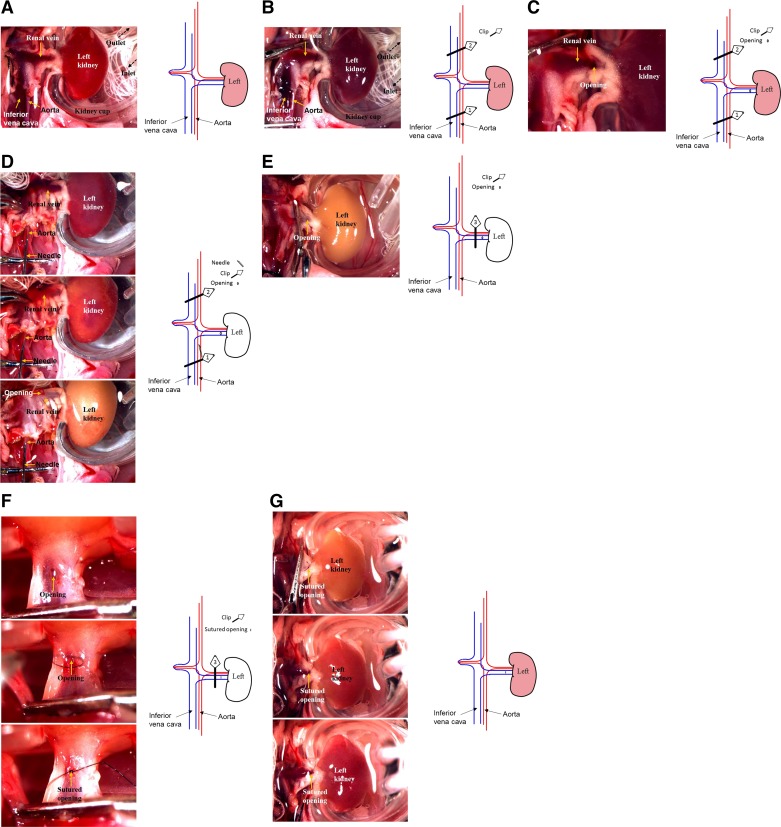
Fig. 1.
Typical procedure images and schematic illustration of the cold ischemia-reperfusion injury (IRI) model. A: the left kidney was placed in a kidney cup and incubated with 4°C saline. B: the inferior vena cava and aorta below and above the renal arteries were clamped (clips 1 and 2, respectively). C: a small opening was cut on the left renal vein. D: the aorta between the left renal artery and clip 1 was injected with sterilized saline using a needle until the color of the left kidney became white. E: the left renal pedicle between the opening and the aorta was clamped (clip 3), and the two clips (clips 1 and 2) on the vena cava and aorta were removed. F: the small opening was sutured. G: renal blood flow was restored.

