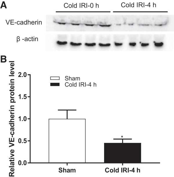
Fig. 5.

Protein level of vascular-endothelial (VE-)cadherin. A: immunoblots of VE-cadherin and the loading control of β-actin for protein normalization. B: protein level of VE-cadherin in the kidney was significantly decreased in the cold IRI-4 h group compared with the cold IRI-0 h group. n = 4. *P < 0.01 vs. the cold IRI-0 h group.
