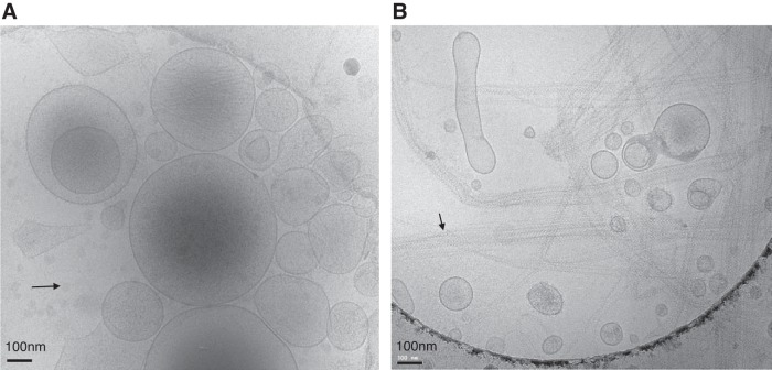
Fig. 2.
Cryo-electron microscopy of extracellular vesicles (EVs) from platelet-poor plasma (A) and urine (B) from two healthy controls (Institutional Review Board-approved protocol no. 17192 at the University of Virginia) after enrichment with differential centrifugation (20,000 g), which is one of the most commonly used EV enrichment techniques. Images show a heterogeneous population of small submicron vesicles of various sizes and densities. However, the bilipid layer morphology is the same for EVs from both body fluids. Of note, EV isolation procedures such as differential centrifugation (e.g., 20,000 g, as used here) can also lead to co-sedimentation and enrichment of non-EV proteins or lipids. The black arrow in A indicates some lipid droplets without a lipid bilayer in the plasma sample. The black arrow in B indicate polymers of Tamm-Horsfall proteins in the urine sample, which are also coprecipitated and may entrap some EVs.

