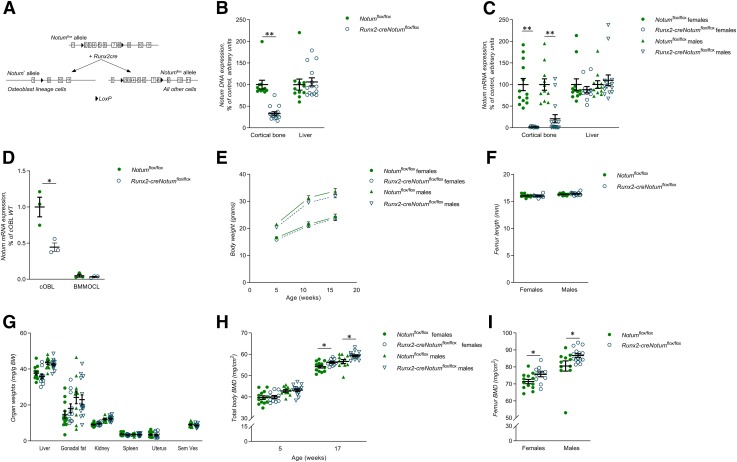
Figure 2.
Osteoblast-derived NOTUM is the principal source of NOTUM in bone. A) Schematic drawing of the conditional osteoblast lineage–specific Notum-inactivated mouse model. B) Degree of deletion of Notum DNA in cortical bone and liver in Runx2-creNotumflox/flox (n = 15) and Notumflox/flox (n = 11) male mice. C) mRNA expression analyses of Notum in the cortical bone and liver in Runx2-creNotumflox/flox (females, n = 11; males, n = 15) and Notumflox/flox (females, n = 13; males, n = 11) mice. D) mRNA expression analyses of Notum in cultured calvarial osteoblasts (cOBL) and bone marrow macrophage–derived osteoclasts (BMMOCLs) from Runx2-creNotumflox/flox and Notumflox/flox mice. (Representative experiment, n = 3 wells/cell type and mouse strain.) E) Normal body weight in Runx2-creNotumflox/flox mice (female, n = 11; male, n = 15) compared to Notumflox/flox mice (female, n = 13; male, n = 11) at 5, 12, and 17 wk of age. F) Normal femur length in Runx2-creNotumflox/flox mice (female, n = 11; male, n = 15) compared to Notumflox/flox mice (female, n = 13; male, n = 11). G) Soft tissue weights over body weight in Runx2-creNotumflox/flox mice (females, n = 11; males, n = 15) and Notumflox/flox mice (females, n = 13; males, n = 11). H) Total body BMD as measured by DXA in 5-wk-old and 17-wk-old Runx2-creNotumflox/flox mice (female, n = 11; male, n = 15) and Notumflox/flox mice (female, n = 13; male, n = 11). I) Femur BMD as measured by DXA in 17-wk-old Runx2-creNotumflox/flox mice (female, n = 11; male, n = 15) and Notumflox/flox mice (female, n = 13; male, n = 11). Unless otherwise stated, the results refer to 17-wk-old mice. All values are given as means ± sem. *P < 0.05, **P < 0.01 vs. Notumflox/flox mice (Student’s t test).

