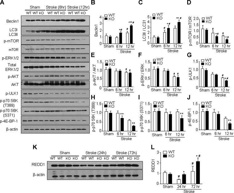
Figure 2.
Increased autophagy in GPR37 KO mice after stroke associated with inhibited mTOR signaling. Western blot analysis was performed to measure several autophagy-related genes in the cortices of sham-treated controls and the peri-infarct region at 6 and 12 h after ischemic stroke. A) Representative Western blots with the indicated antibodies. B) Quantified results showed significant increases in relative Beclin-1 expression in GPR37 KO brain vs. WT control at 6 and 12 h after stroke [2-way ANOVA; F (2,9) = 72.63 l; n = 3/group]. *P < 0.05, ***P < 0.001 vs. WT controls, #P < 0.05 vs. sham-treated control. C) Greater increase in the conversion of LC3-I to LC3-II (larger ratio of LC3-II to LC3-1) after stroke was detected in GPR37 KO mice than that in WT control group [2-way ANOVA; F (2,12) = 95.22; n = 3/group]. *P < 0.05, **P < 0.0001, #P < 0.05 vs. sham-treated control. D) Phosphorylation of the autophagy inhibitory mTOR was significantly suppressed in GPR37 KO mice after stroke [2-way ANOVA; F (2,18) = 47.95; n = 3]. *P < 0.05, #P < 0.05 vs. sham-treated control. E) Phosphorylated AKT showed a trend of reduction in GPR37 KO mice after stroke [2-way ANOVA; F (2,9) = 7.346; n = 3]. #P < 0.05 vs. sham-treated control. F) Phosphorylation of ERK1 and 2 was significantly decreased in both WT and GPR37 KO mice at 12 h after stroke [2-way ANOVA; F (2,9) = 34.73; n = 3]. #P < 0.05 vs. sham-treated control. G) Levels of phosphorylated ULK1 were significantly decreased after stroke in GPR37 KO mice compared with WT mice [2-way ANOVA; F (2,9) = 180.7; n = 3/group]. *P < 0.05 compared with WT stroke controls, #P < 0.05 vs. sham-treated control. H, I) Western blotting of REDD1, an inhibitory factor on the mTOR pathway in the sham-treated and ischemic cortex. There was a transient down-regulation of this factor at 24 h after stroke in WT mice, whereas REDD1 expression was significantly enhanced at 72 h after stroke [F (2,9) = 24.18; n = 3/group]. *P < 0.05 vs. WT, #P < 0.05 vs. sham-treated control.

