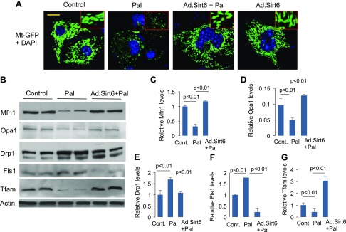
Figure 4.
Sirt6 blocks Pal-induced mitochondrial fragmentation. A) Confocal images of representative cardiomyocytes overexpressed with Ad.Sirt6 and treated with Pal for 24 h. Mitochondrial population was visualized by overexpressing cells with Ad.Mt-GFP vector (green). Position of nuclei was identified by DAPI staining (blue). Inset displays mitochondrial morphology in enlarged image. Scale bar, 2 µm. B) Cell lysate of cardiomyocytes was analyzed by Western blotting using protein-specific antibodies. C–G) Quantitation of the Western blots. Mean ± se; n = 3. Cont., control.

