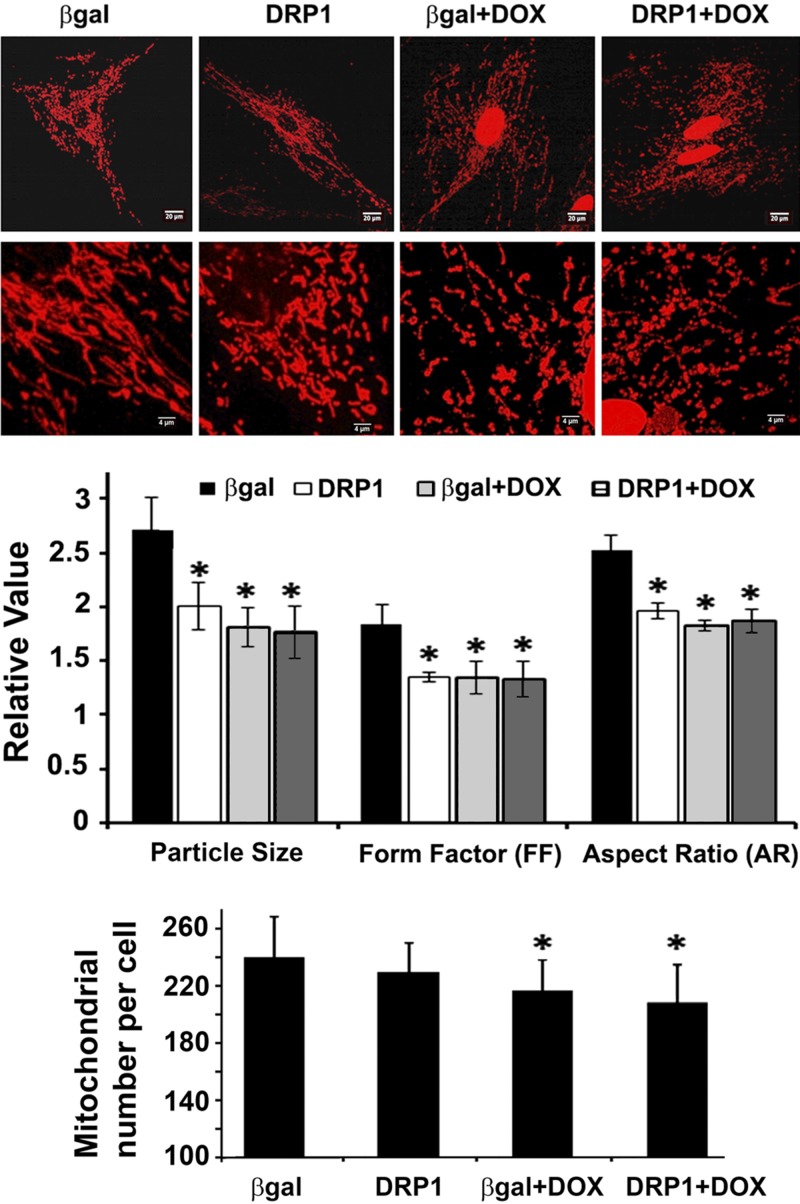
Figure 4.
DRP1 overexpression did not aggravate Dox-induced mitochondrial fragmentation. H9c2 cells were infected with AdDsRed for 48 h, followed by the infection of AdDRP1 or adenovirus expressing β-galactosidase (Adβ-gal) for 48 h. The cells were then exposed to either Dox (750 nM) or saline for 24 h. Mitochondrial morphology was observed with confocal microscopy and analyzed using ImageJ. The following parameters were calculated: mitochondrial particle number and size, mean FF, and AR. At least 5 images (each containing 5–15 cells) were captured per treatment over 3 separate experiments. Data are expressed as means ± se and were analyzed by 1-way ANOVA. *P < 0.05 vs. β-gal (n = 3).

