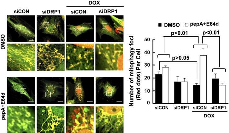
Figure 9.
DRP1 knockdown inhibited Dox-induced mitophagy flux. H9c2 cells were infected with the Admt-Rosella for 48 h. The cells were then transfected with siDRP1 or control siRNA (siCon) for 24 h and exposed to either Dox (750 nM) or saline for 24 h. Confocal images were captured, mitophagy was analyzed using ImageJ, and the number of mitophagy foci per cell was calculated. Between 3 and 8 images (totaling between 5 and 15 cells) were captured per treatment over 3 separate experiments. Scale bars, 20 µm. For evaluating mitophagy flux, experiments were repeated with lysosomal inhibitors (PepA and E64D) or DMSO 4 h after Dox treatment. Data are expressed as means ± se and were analyzed by 1-way ANOVA (P < 0.01 vs. siCon or siCon+Dox; n = 3).

