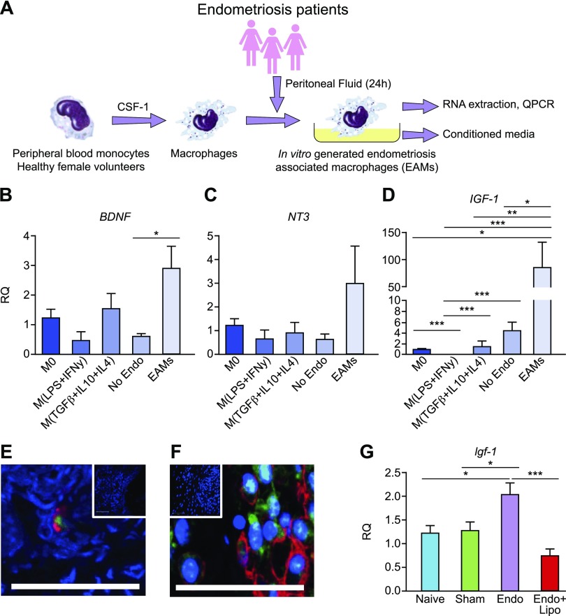
Figure 4.
Disease-modified macrophages in endometriosis exhibit elevated expression of IGF-1. A) Schematic showing generation of EAMs: peripheral blood monocytes from healthy female volunteers (n = 7) were differentiated into macrophages for 7 d in the presence of recombinant M-CSF 1. Macrophages were then activated for 24 h with PF from patients with endometriosis (n = 7) or without endometriosis (n = 8) or activated with different cytokines to generate inflammatory (LPS + IFN-γ) or repair (TGF-β + IL-10 + IL-4; all n = 7) macrophages and compared with M0s. B–D) qPCR revealed that EAMs had significantly increased mRNA concentration of BDNF (P < 0.05) (B), elevated levels of NT-3 (C), and significantly increased concentrations of IGF-1 (P < 0.001) (D). E) Dual immunofluorescence for CD68 (macrophages; red) and IGF-1 (green) revealed that macrophages in human endometriosis lesions express IGF-1. F) In lesions recovered from mice with induced endometriosis, we identified F4/80+ macrophages (red) that stained positive for Igf-1 (green). Scale bar, 50 μM. Top insets show negative controls where the primary antibody is omitted. G) In peritoneal biopsies recovered from endometriosis mice (n = 10), qPCR analysis revealed that mRNA concentration of Igf-1 was elevated (P < 0.05) compared with naive (n = 5) and sham-treated (n = 6) animals. Macrophage depletion in endometriosis mice (n = 10) significantly reduced Igf-1 expression (P < 0.001). RQ, relative quantification. *P < 0.05, **P < 0.01, ***P < 0.001.

