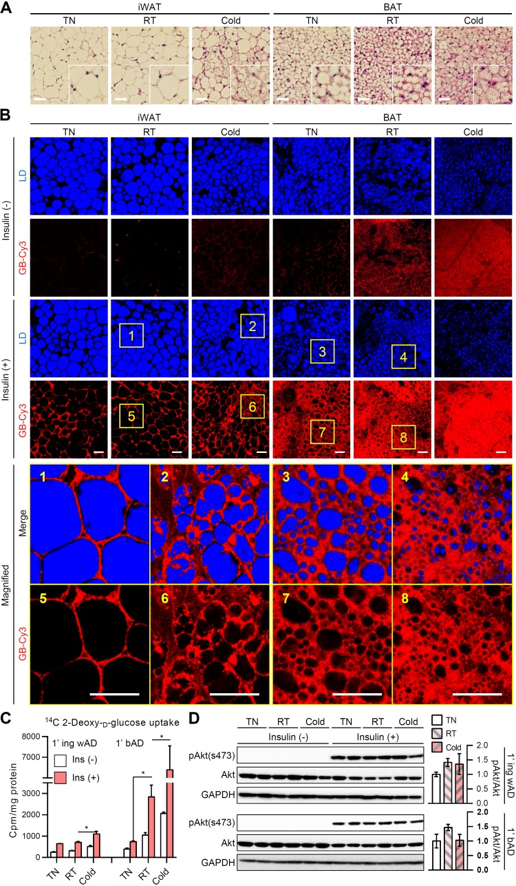
FIG 1.
Differential regulation of insulin-dependent glucose uptake in adipocytes upon different temperatures. Eight-week-old male C57BL/6J mice were placed under thermoneutral (30°C), room temperature (25°C), or cold (4°C) conditions for 1 week. (A) Hematoxylin-eosin-stained paraffin sections of inguinal white adipose tissue (iWAT) and brown adipose tissue (BAT). Insets show ×2 magnifications of the indicated areas. Scale bars, 50 μm. (B) Ex vivo insulin-dependent glucose bioprobe (GB-Cy3, red) uptake assay. iWATs and BATs from mice were cultured with GB-Cy3 (5 μM) in the absence or presence of insulin (1 μM, 30 min). Lipid droplets (LDs, blue) were visualized by TCS SP8 CARS microscopy. Scale bars, 50 μm. Insets show ×5 magnifications of the indicated areas. (C and D) Insulin-dependent glucose uptake (C) and immunoblot of insulin signaling cascade (D) in primary adipocytes isolated from iWAT (1′ ing wAD) and BAT (1′ bAD) with or without insulin (100 nM, 30 min). Data represent means ± the standard deviations (SD). *, P < 0.05 (Student t test).

