FIG 6.
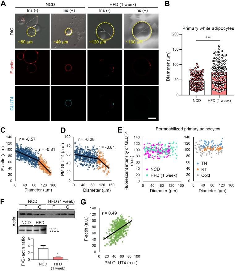
In WATs, obesity-induced LD enlargement suppress insulin-dependent GLUT4 translocation to the PM. Primary white adipocytes were isolated from eWATs of 8-week-old male C57BL/6J mice fed the HFD for 1 week. (A) F-actin (phalloidin-TRITC, red) and PM-anchored GLUT4 (cyan) of primary white adipocytes were assessed by immunohistochemistry in the absence or presence of insulin (100 nM, 30 min). Scale bar, 50 μm. (B) The diameters of isolated primary white adipocytes were quantified using ImageJ. (C and D) Size-dependent fluorescent intensities of PM-anchored GLUT4 (C) and F-actin (D). (E) Total GLUT4 fluorescence intensities were measured in primary adipocytes after permeabilization. (F) F/G-actin ratios. WCL, whole-cell lysates. (G) Fluorescence intensities of F-actin and PM-anchored GLUT4. r, correlation coefficient. Data represent means ± the SD. *, P < 0.05 and ***, P < 0.001 versus NCD (Student t test).

