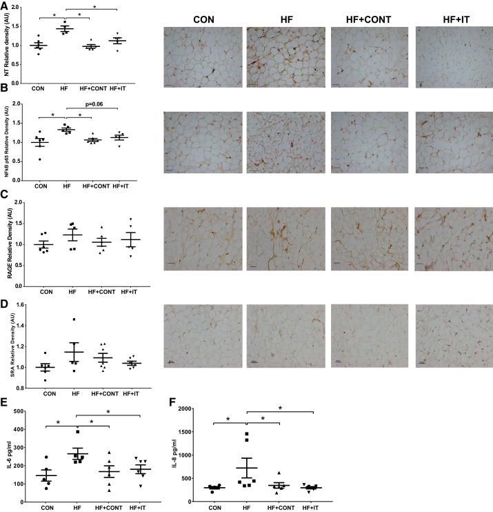
Fig. 6.
Immunohistochemistry analysis of oxidative stress and inflammation in swine pericoronary adipose tissue and cytokine levels in swine perivascular adipose tissue (PVAT) conditioned medium. A and B: greater nitrotyrosine (NT; A) and NF-κB p65 (B) in heart failure (HF) was attenuated by both HF+CONT and HF+IT. CON, control; HF+CONT, HF continuous exercise trained; HF+IT, HF interval exercise trained. C and D: receptor of advanced glycation end products (RAGE; C) and scavenger receptor A (SRA; D) levels were unchanged in all groups. AU, arbitrary units. E and F: IL-6 (E) and IL-8 (F) secretion in HF conditioned medium was prevented by HF+CONT and HF+IT (n = 6/group). Data are means ± SE.*HF vs. CON, HF+CONT, and HF+IT, P < 0.05. Data were analyzed with a 1-way ANOVA. Right: representative immunohistochemistry images of coronary artery showing pericoronary adipose tissue expression of NT, NF-kB p65, RAGE and SRA. Scale bars, 100 μm.

