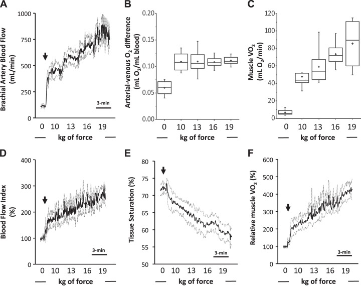
Fig. 1.
A–C: conventional measures of muscle O2 consumption. A: Doppler-derived brachial artery blood flow. B: arterial-venous O2 difference. C: skeletal muscle O2 consumption (V̇o2). D–F: diffuse correlation spectroscopy (DCS)-derived determinants of relative muscle O2 consumption.D: DCS-derived blood flow index. E: near-infrared spectroscopy-derived skeletal muscle tissue saturation. F: relative muscle V̇o2. Rhythmic handgrip began with a fixed workload of 10 kg and progressed by 3 kg every 3 min thereafter. Arrows depict the onset of exercise. Data are reported as means ± SE; n = 8.

