Fig. 1.
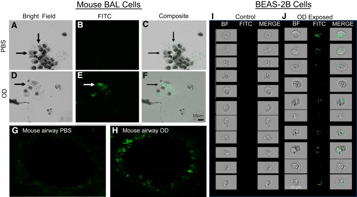
Epithelial cells engulf organic dust (OD) in vivo and in vitro. A–C :unstained bronchoalveolar lavage (BAL) cells from PBS-treated mice were observed under light microscopy (A), fluorescence (B), and a composite (C). D: BAL cells from OD-exposed mice under bright field (BF). E: under fluorescence, OD is observed in bronchial epithelial cells. F: composite of bright field and fluorescent channel confirmed the presence of OD in bronchial epithelial cells from OD mice. G: an airway from control-treated mouse, showing no autofluorescence present in bronchial epithelial cells. H: airway from OD-treated mouse, with a high degree of fluorescence localized to bronchial epithelial cells. I: BEAS-2B cells exposed to PBS for 24 h. J: BEAS-2B cells exposed to 100 μg OD for 24 h showed autofluorescent OD intracellularly using ImageStreamX.

