Fig. 2.
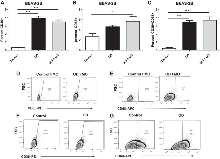
Organic dust (OD) exposure increases phagocytic markers in BEAS-2B cells. Phagocytic marker expression was assessed after 24 h in OD-exposed cells with or without sulforaphane (Sul) pretreatment. A: increase in the percentage of CD36+ cells after OD or Sul + OD compared with control. B: increase in the percentage of CD68+ cells after OD or Sul + OD compared with control. C: the number of positive cells expressing both CD36 and CD68 was increased after OD exposure compared with control cells. D and E: gating strategies for the CD68 and CD36 are shown. F and G: representative plots showing increase in markers before and after OD. All data were analyzed using ANOVA. *P < 0.5, ***P < 0.001; n = 4 independent experiments. APC, allophycocyanin; FMO, fluorescence minus one; FSC, forward scatter; PE, phycoerythrin.

