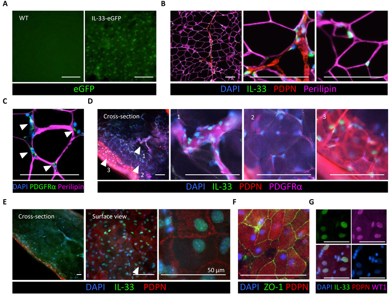
Fig. 3.
Localization of IL-33-expressing cells in visceral WAT. (A) Surface view of the ewat from the IL-33-eGFP reporter mouse (right) and a wild type (WT) control animal (left). (B) Immunostaining of IL-33 (green) in paraffin-embedded WT mouse ewat. Note IL-33 localization to DAPI-stained nuclei (blue) scattered between perilipin+ adipocytes (purple), as well as IL-33 in the PDPN+ (red) membrane lining the adipose compartment. (C) Immunostaining of paraffin-embedded WT mouse ewat. PDGFRα-expressing ASPCs (green) embedded between perilipin+ adipocytes (purple). (D) Immunostained transverse sections (side view) of whole-mount WT mouse ewat demonstrating PDGFRα-stained (purple) ASPCs that express IL-33 (green) in the nucleus (blue). The PDPN-stained (red) cell layer expressing nuclear IL-33 (green) envelops the adipose compartment. Arrowheads indicate magnified areas in adjacent panels. (E) Side and surface view of the same sample of WT mouse whole-mount stained ewat. (F and G) Surface view of whole-mount stained ewat. ZO1 (green) staining on PDPN+ (red) cobblestone-shaped cells in (F) and co-localization of IL-33 (green) and WT1 (purple) in the nucleus (blue) in (G). Scale bar, 100 μm, unless indicated otherwise. All immunostaining experiments are representative of two mice, and the staining protocol has been repeated minimum of three times.

