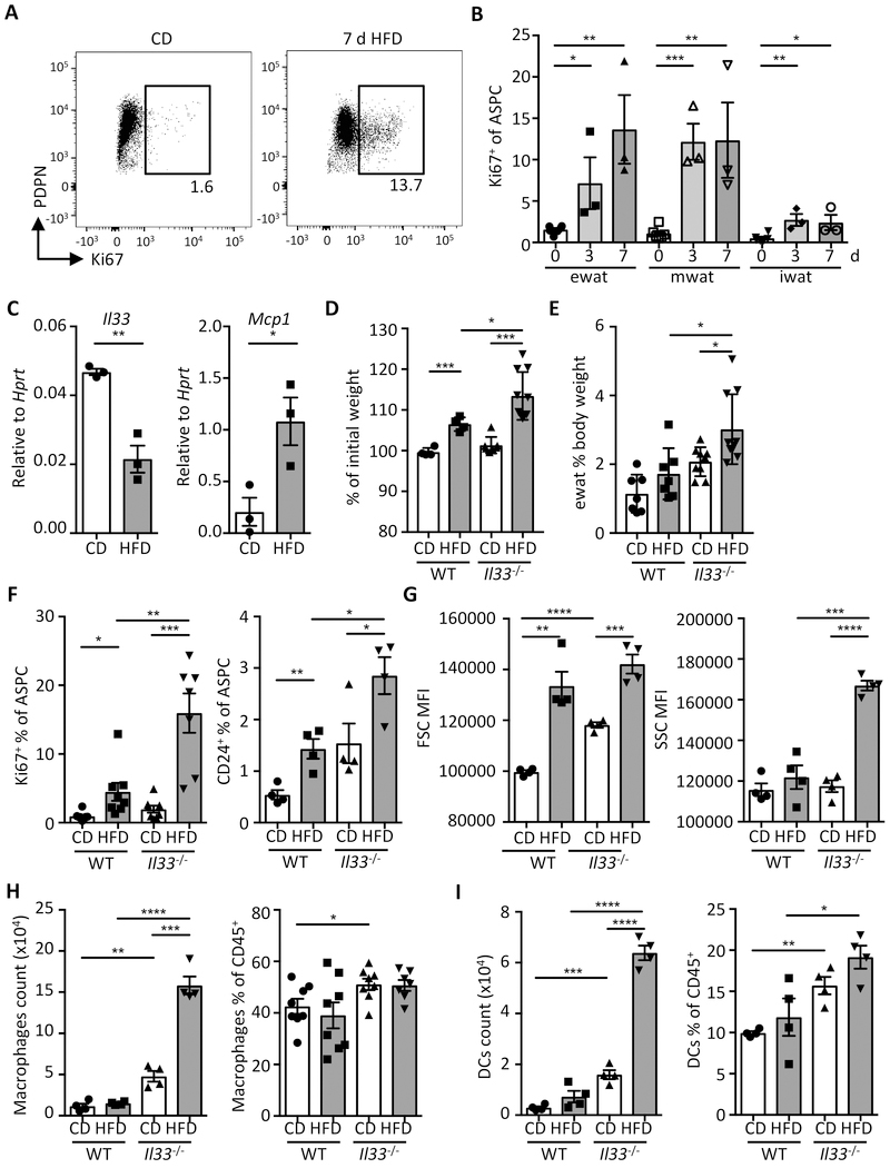
Fig. 5.
IL-33 controls adipose tissue expansion and immunological homeostasis in short-term HFD feeding. (A-B) Wild type mice were fed with high-fat diet (HFD) or control diet (CD) up to 7 days (d). (A) Representative dot plot of Ki67 staining in ASPCs (defined as live lin− (CD31− CD45− Ter119−)) PDGFRα+Sca-1+ cells) on day 7. (B) Proportion of proliferating cells (Ki67+) cells among total ASPCs from indicated WAT depots. (n = 3) (C) Expression of Il33 and Mcp1 in sort-purified ASPCs isolated from ewat of wild type mice (WT) on CD or HFD for 3 days as determined by qRT-PCR. (n = 3) (D to I) WT or Il33−/− mice were fed with CD or HFD and sacrificed for analysis after 3 days. (D) Body weight change as a proportion (%) of initial weight (day 0). Data compiled from two individual experiments. (n ≥ 7) (E) Proportion of ewat mass as proportion (%) of body weight. Data compiled from two individual experiments. (n ≥ 7) (F) Proportion of Ki67+ and adipocyte progenitor cells (CD24+) among total ewat ASPCs. (G) Mean fluorescence intensity (MFI) of forward scatter (FSC) and side scatter (SSC) of ewat ASPCs. (H and I) Quantification of macrophages (H) and dendritic cells (DCs) (I) in ewat. (n ≥ 4). Mean ± SEM, * P < 0.05, ** P < 0.01, ***P < 0.001, Student’s t-test.

