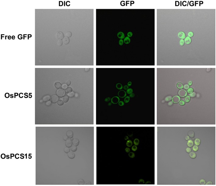
Figure 5.

Subcellular localisations of OsPCS5 and OsPCS15 proteins tagged with GFP in yeast cells. Saccharomyces cerevisiae DTY167 cells were transformed with free GFP, OsPCS5::GFP or OsPCS15::GFP, and observed via confocal fluorescence microscopy. DIC and GFP merged images were generated using Olympus software. DIC, differential interference contrast; GFP, green fluorescent protein; DIC/GFP, merged images of DIC and GFP.
