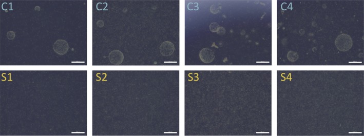
Figure 2.

Light micrographs of colonial and solitary culture replicates immediately prior to RNA extraction. Each colonial (C1–C4) and solitary (S1–S4) replicate was imaged with an Olympus CKX53 light microscope just before harvesting cells for RNA extraction. Colonies were visible in all colonial replicates and absent in all solitary replicates. Colonial replicate C2 had a high density of solitary cells compared to the other colonial replicates at the time of RNA extraction. Scale bars are 500 μm in all images.
